Muscle Contraction - Part 2 Summary Questions
- Due Apr 21, 2020 at 11:59pm
- Points 12
- Questions 12
- Time Limit None
- Allowed Attempts 3
Instructions
Be able to answer this question on the next page:
- Explain the steps to an action potential.
10.3 | Muscle Fiber Contraction and Relaxation
By the end of this section, you will be able to:
- Describe the components involved in a muscle contraction
- Explain how muscles contract and relax
- Describe the sliding filament model of muscle contraction
From Part 1: (To review)
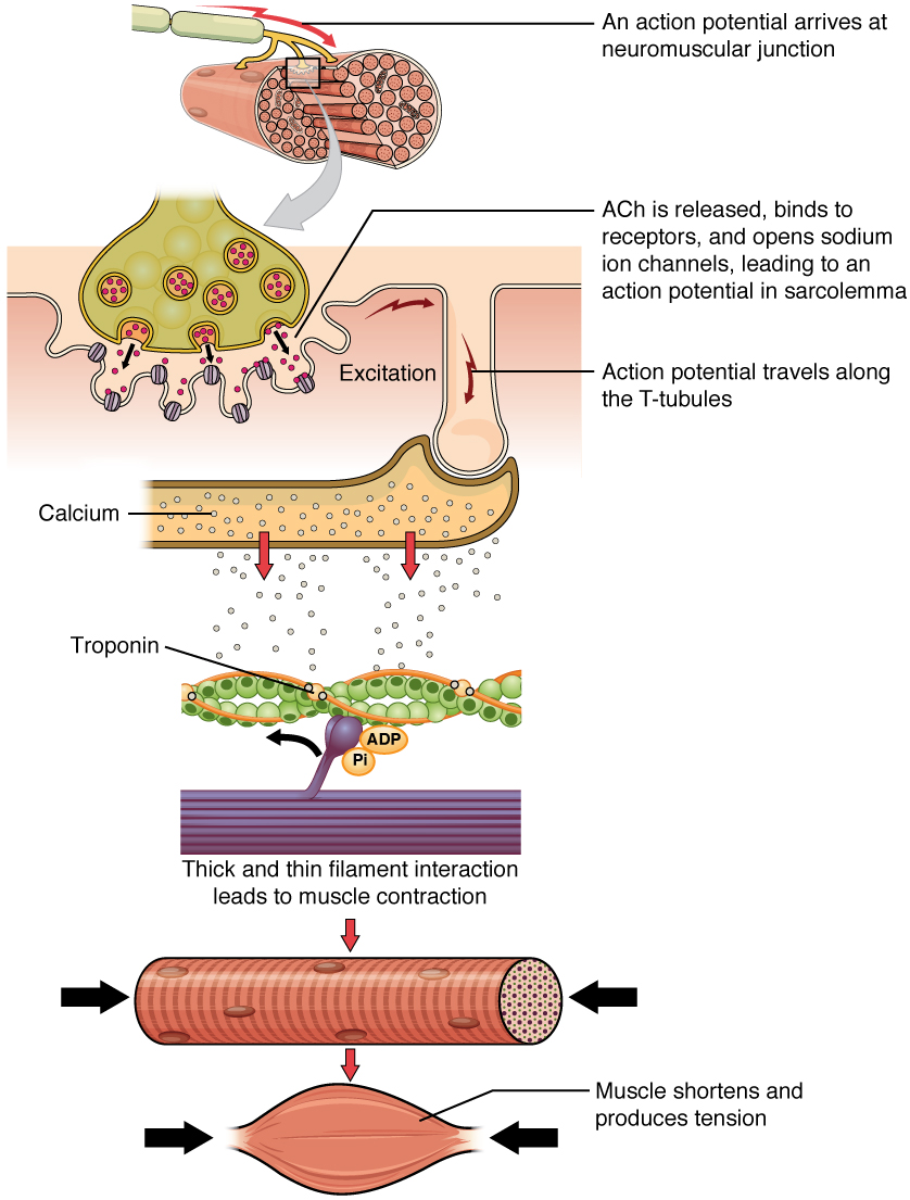
The sequence of events that result in the contraction of an individual muscle fiber begins with a signal—the neurotransmitter, ACh—from the motor neuron innervating that fiber.
- The local membrane of the fiber will depolarize as positively charged sodium ions (Na+) enter, triggering an action potential that spreads to the rest of the membrane will depolarize, including the T-tubules.
- This triggers the release of calcium ions (Ca++) from storage in the sarcoplasmic reticulum (SR).
Starting Part 2:
- The Ca++ then initiates contraction, which is sustained by ATP.
- As long as Ca++ ions remain in the sarcoplasm to bind to troponin, which keeps the actin-binding sites “unshielded,” and as long as ATP is available to drive the cross-bridge cycling and the pulling of actin strands by myosin, the muscle fiber will continue to shorten to an anatomical limit.

Contraction of a muscle fiber: A cross-bridge forms between actin and the myosin heads triggering contraction. As long as Ca++ ions remain in the sarcoplasm to bind to troponin, and as long as ATP is available, the muscle fiber will continue to shorten.
Muscle contraction usually stops when signaling from the motor neuron ends, which repolarizes the sarcolemma and Ttubules, and closes the voltage-gated calcium channels in the SR.
- Ca++ ions are then pumped back into the SR, which causes the tropomyosin to reshield (or re-cover) the binding sites on the actin strands.
- A muscle also can stop contracting when it runs out of ATP and becomes fatigued.
Relaxation of a muscle fiber: Ca++ ions are pumped back into the SR, which causes the tropomyosin to reshield the binding sites on the actin strands.
- A muscle may also stop contracting when it runs out of ATP and
becomes fatigued.
The release of calcium ions initiates muscle contractions.
- Watch this video to learn more about the role of calcium.https://www.youtube.com/watch?v=CepeYFvqmk4
The molecular events of muscle fiber shortening occur within the fiber’s sarcomeres.
- The contraction of a striated muscle fiber occurs as the sarcomeres, linearly arranged within myofibrils, shorten as myosin heads pull on the actin filaments.
The region where thick and thin filaments overlap has a dense appearance, as there is little space between the filaments.
- This zone where thin and thick filaments overlap is very important to muscle contraction, as it is the site where filament movement starts.
- Thin filaments, anchored at their ends by the Z-discs, do not extend completely into the central region that only contains thick filaments, anchored at their bases at a spot called the M-line.
- A myofibril is composed of many sarcomeres running along its length; thus, myofibrils and muscle cells contract as the sarcomeres contract.
The Sliding Filament Model of Contraction
When signaled by a motor neuron, a skeletal muscle fiber contracts as the thin filaments are pulled and then slide past the thick filaments within the fiber’s sarcomeres.
- This process is known as the sliding filament model of muscle contraction.
- The sliding can only occur when myosin-binding sites on the actin filaments are exposed by a series of steps that begins with Ca++ entry into the sarcoplasm.

The Sliding Filament Model of Muscle Contraction - When a sarcomere contracts, the Z lines move closer together, and the I band becomes smaller.
- The A band stays the same width.
- At full contraction, the thin and thick filaments overlap.
Tropomyosin is a protein that winds around the chains of the actin filament and covers the myosin-binding sites to prevent actin from binding to myosin.
- Tropomyosin binds to troponin to form a troponin-tropomyosin complex. The troponintropomyosin complex prevents the myosin “heads” from binding to the active sites on the actin microfilaments.
- Troponin also has a binding site for Ca++ ions.
To initiate muscle contraction, tropomyosin has to expose the myosin-binding site on an actin filament to allow cross-bridge formation between the actin and myosin microfilaments.
- The first step in the process of contraction is for Ca++ to bind to troponin so that tropomyosin can slide away from the binding sites on the actin strands.
- This allows the myosin heads to bind to these exposed binding sites and form cross-bridges.
- The thin filaments are then pulled by the myosin heads to slide past the thick filaments toward the center of the sarcomere.
- But each head can only pull a very short distance before it has reached its limit and must be “re-cocked” before it can pull again, a step that requires ATP.
HERE IS YOUR SUMMARY AND WHAT YOU WILL NEED TO KNOW FOR THE QUIZ NEXT TUESDAY.
-
- YOU WILL ALSO NEED TO KNOW THE NAMES OF YOUR MUSCLES.
Part 1 and Part 2:
- The nervous system sends a signal through a nerve to the muscle.
- Ach neurotransmitter is released into the synaptic cleft from the nerve.
- Ach binds to the receptor and triggers it to open allowing Na+ to enter cell
- Depolarization occurs and the action potential (excitation) is started in the muscle.
- Exciting of the membrane triggers Ca+ to be released from the SR (sarcoplasmic reticulum) into the sarcoplasm (by using T-tubules)
- Ca+ binds with the shielding proteins troponin.
- Tropomyosin slide away from the actin binding sites.
- The actin binding sites are unshielded allowing myosin to attach to actin.
- Myosin pulls the actin filaments toward center to shorten the muscle.
- Muscle contraction stops when Ach is degraded from the synaptic cleft.
- Na+ is pumped out of the cell and the membrane is repolarized with a negative charge on the inside.
- Ca+ is taken back into the SR and troponin will rebind to actin.